


3D-printed scaffolds that mimic the chemical composition and architecture of natural bone tissue (Image courtesy Jonne Renvall, Tampere University)
Bone grafting is the second most common tissue transplantation procedure worldwide, with more than two million operations performed annually. Current treatments often rely on bone taken from the patient or a donor, approaches that are limited in availability and may involve additional surgery, lengthy recovery times and complications. As populations age, the need for safer and more effective alternatives is growing rapidly.
The research led by Antonia Ressler, Postdoctoral Research Fellow at the Tampere Institute for Advanced Study, has successfully used hydroxyapatite, the same compound that forms the mineral structure of natural bone, to create bone-like scaffolds that support the body’s own capacity for tissue regeneration. “By using the same material that nature uses and shaping it through ceramic 3D printing, the implants can be precisely tailored to match a patient’s individual bone defect, without relying on drugs or growth factors that may cause side effects,” says Ressler.
The novel technology is the result of four years of intensive research in the AffordBoneS project funded by the Horizon Europe Marie Skłodowska-Curie Postdoctoral Fellowship programme. An ongoing project, GlassBoneS, aims to further develop this technology. The research team aims to provide affordable scaffolds for bone augmentation procedures to enable broader access to these treatments and improve patients’ quality of life.
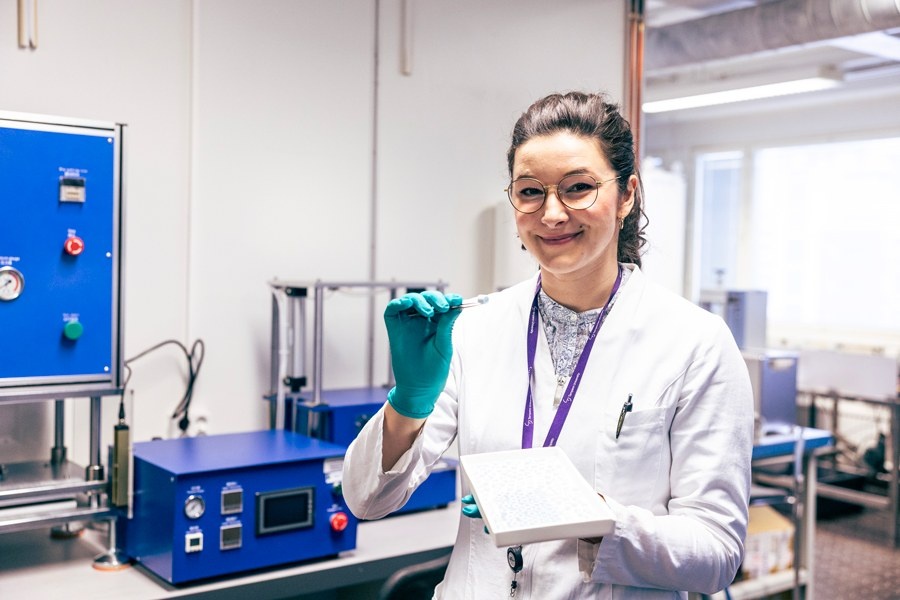
Lead researcher Antonia Ressler (Image courtesy Jonne Renvall, Tampere University)
Individually Designed Bone Grafts
Using an advanced manufacturing technique known as ceramic 3D printing, the researchers were able to precisely control the internal architecture of the scaffolds, including the size and connectivity of pores that allow cells to grow and nutrients to flow through the material. The team identified an optimal bone-like structure: implants with carefully designed internal pores of around 400 micrometres and approximately 45% porosity.
“This architecture achieved a crucial balance between strength and biological performance, allowing bone-forming cells to enter the material, interact with one another, and successfully begin forming new bone tissue,” says Ressler.
The team also discovered that subtle changes in material chemistry and surface properties can influence cell behaviour. “We found that the high temperatures required during processing can alter the surface of the material in ways that make it more difficult for human cells to attach. Our finding highlights that not only the composition, but also the surface properties of biomaterials are critical for successful bone regeneration,” she says. (Via Tampere University)
Source:
https://www.sciencedirect.com/science/article/pii/S2590006426003170
Also read: