Ultrasound can now be used to image single cells inside the body
https://press.asimov.com/articles/gas-vesicles


Ultrasound can now be used to image single cells inside the body
https://press.asimov.com/articles/gas-vesicles
1 Comment
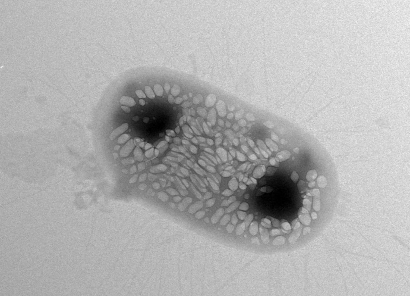
In the 1890s, a German microbiologist discovered gas vesicles, rigid protein structures that trap gas, in a species of cyanobacteria called Gleotrichia echinulata. But it wasn’t until the 1960s that scientists began to study gas vesicles in more detail, eventually revealing that they could reflect sound waves in a way that tissues in the body cannot, making them visible on ultrasound scans.
In just the last few years, scientists have engineered gas vesicles and inserted them into bacteria and human cells. They can then use ultrasound to image cells containing these gas vesicles with amazing resolution. In other words, scientists can now watch cells move through the body in a way that microscopy, MRI, and other methods cannot.
But when will these be used in clinical trials, if ever? What are their actual applications?